Intrinsic vs. Extrinsic Teeth Stains: Causes & Clinical Removal Guide
October 23, 2026
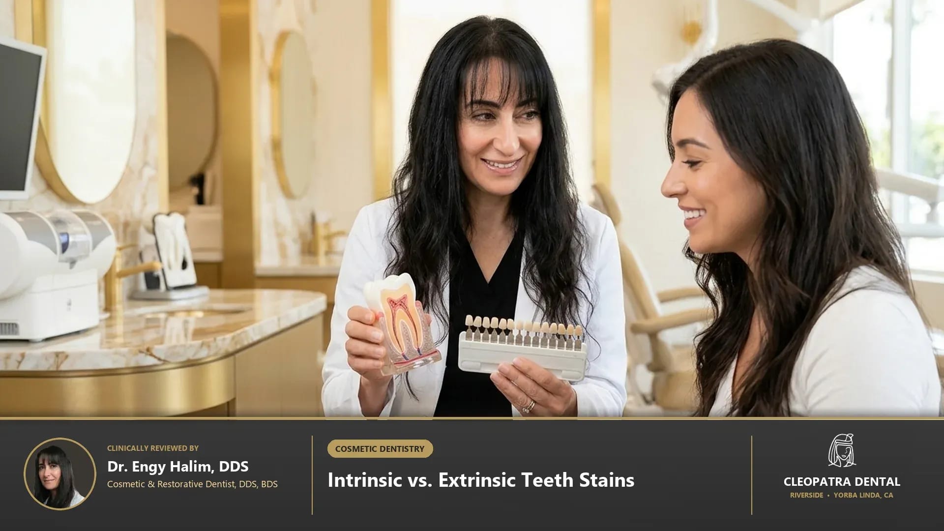
Quick Verified Summary: Extrinsic teeth stains accumulate on the outer enamel pellicle from chromogenic coffee, tea, red wine, tobacco, and chlorhexidine, responding rapidly to ultrasonic prophylaxis (CDT D1110) and peroxide whitening (D9972). Intrinsic stains originate inside the underlying dentin from tetracycline antibiotics, dental fluorosis, pulp necrosis, or aging, requiring internal bleaching (D9974) or IPS e.max porcelain veneers (D2962).
Clinical Foundations: Understanding Intrinsic vs. Extrinsic Teeth Stains: Causes & Clinical Removal Guide
Patients researching extrinsic and intrinsic stains in Southern California deserve transparent, evidence-based guidance rooted in biological tooth preservation and long-term oral systemic health. At Cleopatra Dental, our general, restorative, and surgical clinicians evaluate every patient as a complete biomechanical system rather than treating isolated symptoms. In daily practice across Western Riverside County and North Orange County, we find that differential shade diagnosis under natural D65 daylight illumination (5,500K), transillumination, and periapical radiography distinguishes superficial salivary pellicle chromogens from internal dentinal chelates and pulpal hemorrhage. This diagnostic precision prevents guesswork, minimizes chairside discomfort, and establishes predictable clinical outcomes for families and older adults alike. Patients looking to erase stubborn enamel chromogens can book professional in-office and custom tray teeth whitening at our practice.
Modern dentistry has evolved rapidly away from aggressive mechanical preparation toward minimally invasive, biologically guided care. When evaluating intrinsic vs extrinsic teeth stains, our diagnostic team examines how enamel thickness, underlying dentin tubules, periodontal ligament fibers, and occlusal biting forces interact over a 10- to 20-year horizon. By integrating digital optical scanning, low-dose imaging, and biocompatible materials coded under ADA CDT standards (D9972, D9974, D2962, D1110), we tailor each treatment plan to your specific anatomy, medical history, and comfort preferences. When deep developmental or medication-induced dentin banding resists bleaching, we craft custom IPS e.max porcelain veneers to permanently mask discoloration.
Biological & Biomechanical Principles Behind intrinsic vs extrinsic teeth stains
Every clinical decision in restorative and preventive dentistry depends on respecting microscopic tissue thresholds. Scientific studies confirm that hydrogen peroxide (H2O2, molecular weight 34.01 g/mol) diffuses through interprismatic enamel micropores into the dentinoenamel junction (DEJ), releasing free-radical oxygen species that cleave double-bonded carbon rings in dark organic chromophores into colorless, shorter-chain molecules. When these quantitative parameters are carefully controlled at the chairside, natural dental tissues and prosthetic restorations withstand daily mastication forces—which routinely reach 70 to 150 pounds per square inch during normal chewing and exceed 250 pounds per square inch during nocturnal clenching—without premature wear, sensitivity, or structural fatigue. Compare clinical peroxide concentrations for lifting stubborn chromophores in our guide to custom teeth whitening trays vs. in-office Opalescence Boost.
Advanced Biomaterials and Digital Diagnostic Standards
Material science plays an equally decisive role in clinical longevity and biocompatibility. In our clinical protocols, we utilize ultrasonic piezoelectric scaling + glycine/erythritol air polishing (CDT D1110), 40% pH-neutral hydrogen peroxide chairside gel (D9972), 10%–16% carbamide peroxide custom trays (D9975), sodium perborate walking bleach for non-vital root-canal teeth (D9974), and 0.3 mm lithium disilicate veneers (D2962). Unlike legacy dental materials that relied on mechanical wedging or high-expansion metals, contemporary ceramic, polymer, and biologic systems bond adhesively or integrate directly with host tissues. This seals out cariogenic oral bacteria, maintains neutral salivary pH balance, and delivers natural optical translucency that blends seamlessly with your surrounding smile. For deep tetracycline or developmental stains that resist bleaching, learn about minimal-prep porcelain veneers for intrinsic tooth stains.
Side-by-Side Clinical Comparison & Candidacy Matrix
Choosing the right clinical pathway requires comparing treatment modalities across biological conservation, expected service life, patient comfort, and maintenance requirements. During your consultation, our doctors review high-resolution intraoral photographs with you on a chairside monitor so you can see exactly what we see. The clinical comparison matrix below summarizes how each diagnostic and therapeutic option performs in real-world practice. See how different bleaching systems rank by shade lift and Relative Dentin Abrasivity in which tooth whitening system works best for enamel safety.
| Stain Classification & Subtype | Histological Location & Molecular Cause | Visual Color & Pattern Presentation | Definitive Clinical Treatment Protocol (ADA CDT) |
|---|---|---|---|
| Direct Extrinsic Dietary Stains | Adsorbed onto acquired salivary enamel pellicle via hydrogen/electrostatic bonds | Uniform yellow-brown tint on lingual and interproximal surfaces (coffee, tea, red wine) | Prophylaxis air-polishing (D1110) + 40% H2O2 in-office or custom tray whitening (D9972/D9975) |
| Indirect Extrinsic Cationic/Metallic Stains | Chemical precipitation of cationic antimicrobials or metal salts with dietary tannins | Dark brown/black cervical specks from 0.12% chlorhexidine rinse, iron supplements, or chromogenic bacteria | Ultrasonic scaling and rubber-cup prophylaxis paste polishing by an RDH (D1110) |
| Intrinsic Single-Tooth Non-Vital Stain | Hemoglobin breakdown (iron sulfide hemosiderin) inside dentinal tubules after pulp trauma/root canal | Isolated dark gray, purple, or brown front tooth next to bright natural neighbors | Non-vital intracoronal "walking bleach" with sodium perborate (D9974) or ceramic crown/veneer |
| Intrinsic Tetracycline Chelation Banding | Calcium-orthophosphate tetracycline complex incorporated into calcifying dentin during childhood | Horizontal yellow, gray, or dark blue-brown banding across entire dental arch | Extended 6-to-12-week 10% carbamide peroxide trays (D9975) or IPS e.max porcelain veneers (D2962) |
| Intrinsic Enamel Fluorosis & Hypoplasia | Excess systemic fluoride (> 2 ppm) or high fever during amelogenesis creating subsurface porosity | Chalky opaque white flecks (mild) to pitted brown enamel patches (moderate/severe) | Enamel microabrasion + Icon resin infiltration (D2990) or minimal-prep veneers (D2962) |
How We Customize Candidacy for Complex Medical Histories
No two mouths age or heal identically. Patients managing systemic conditions such as Type 2 diabetes, hypertension, autoimmune disorders, osteoporosis, or medication-induced dry mouth (xerostomia) require customized dental protocols. Before initiating treatment related to intrinsic teeth stains, we review your current medications, allergies, and systemic health markers. Adjusting local anesthetic buffering, selecting hypoallergenic metal-free ceramics, and reinforcing salivary mineral buffering ensures safe, predictable healing for every age group. Older adults experiencing age-related yellowish dentin show-through can read whether teeth whitening is safe for seniors over 60 with thinning enamel.
Preventing Common Complications and Premature Failure
Even the finest dental restoration or preventive regimen requires protection against mechanical overload and bacterial biofilm accumulation. In clinical audits, the two leading causes of preventable dental breakdown are unaddressed nighttime teeth grinding (sleep bruxism) and interproximal plaque stagnation along the gumline. Addressing occlusal harmony early and establishing an easy-to-follow home care routine protects both your natural enamel and any restorative work for decades.
Long-Term Maintenance & Preventive Follow-Up in Southern California
Sustaining healthy clinical results over 10 to 20 years depends on partnering with a consistent dental team that monitors your gum attachment levels, bite balance, and restorative margins at every six-month checkup. At our Riverside and Yorba Linda locations, we combine ultrasonic hygiene care, digital bitewing monitoring, and occlusal nightguard checks to catch micro-changes long before they become costly emergencies.
2026 Cost Guide, ADA CDT Codes & PPO Insurance in Riverside and Yorba Linda
Financial clarity is a core pillar of patient trust at Cleopatra Dental. Treatment fees in Southern California vary according to diagnostic complexity, the number of teeth or quadrants involved, laboratory biomaterial selection, and your dental PPO insurance benefits. Before any procedure begins, our treatment coordinators verify your insurance eligibility and provide a clear, written breakdown organized by official American Dental Association Current Dental Terminology (CDT) codes.
Important Pricing Note: The prices shown in the table below are estimated ranges; to obtain accurate pricing tailored to your specific clinical needs and insurance coverage, please contact our Riverside or Yorba Linda office.
| Stain Removal & Cosmetic Procedure (ADA CDT) | Estimated Cost & Price Range (2026) | Expected Shade Lift & Coverage Notes |
|---|---|---|
| Adult Prophylaxis Cleaning & Stain Polishing (D1110) | $115 – $195 | Removes 100% of superficial extrinsic plaque and tartar stains; 100% PPO preventive coverage |
| Custom Scalloped Take-Home Whitening Trays (D9975) | $295 – $495 | Lifts yellow/brown extrinsic & mild intrinsic stains 4–6 VITA shades over 10–14 days |
| In-Office 40% Hydrogen Peroxide Boost Whitening (D9972) | $450 – $750 | Lifts 6–8 VITA shades in 60 minutes with potassium nitrate desensitizer |
| Internal Bleaching of Non-Vital Endodontic Tooth (D9974) | $350 – $650 | Per tooth; whitens darkened root-canal-treated tooth from the inside out |
| Minimal-Prep IPS e.max Porcelain Veneer (D2962) | $1,250 – $2,400 | Permanent mask for severe Grade III/IV tetracycline or fluorosis stains; CareCredit 0% APR |
Maximizing Dental PPO Insurance, HSA/FSA Funds & 0% Financing
Most PPO dental insurance plans categorize diagnostic exams, digital X-rays, and routine cleanings under preventive benefits covered at 80% to 100%, while basic and major restorative procedures receive 50% to 80% coverage up to your annual plan maximum (typically $1,500 to $2,500 per calendar year). For treatments that exceed annual insurance caps or involve elective cosmetic enhancements, patients can apply tax-free Health Savings Account (HSA) or Flexible Spending Account (FSA) dollars, or enroll in 0% APR promotional monthly financing through CareCredit, LendingClub, and Proceed Finance.
Step-by-Step Clinical Protocol & Long-Term Care Guidelines
Knowing exactly what happens before, during, and after your visit eliminates dental anxiety and empowers you to participate actively in your oral health. Our clinical workflow enforces Supervised under CA B&P § 1907–1926 by Dr. Engy Halim, DDS and Dr. George Mirsal, DDS alongside Tiffany Anderson, RDH; enforcing a 48-hour white diet post-bleaching while hydroxyapatite prisms rehydrate and remineralize with 5% potassium nitrate and 1,100–5,000 ppm fluoride. By breaking complex care into calm, well-paced stages, we ensure your comfort from initial check-in through long-term maintenance. Spectrophotometric VITA Classical and 3D-Master shade assessments are performed daily at our Riverside cosmetic dentistry clinic.
Step 1: Comprehensive Digital Assessment and Shade/Bite Mapping
Your appointment begins with a conversation about your goals, symptoms, and past dental experiences, followed by gentle digital diagnostics. Using radiation-free iTero 3D optical scanning, intraoral high-definition cameras, and ultra-low-dose digital radiographs when indicated, we map your teeth, gums, and bite registration within minutes—eliminating messy impression trays and providing an immediate 3D visual roadmap of your mouth. Orange County patients can also schedule customized stain diagnosis and whitening at our Yorba Linda cosmetic dental studio.
Step 2: Precision Chairside Execution and Post-Care Verification
During active care, we prioritize profound comfort using topical numbing gels, pH-buffered local anesthetics, and optional nitrous oxide or oral conscious sedation. Every margin, contact point, and biting surface is verified under high magnification before you leave the chair. You also receive written post-visit care instructions and a direct follow-up call from our clinical team to confirm your recovery is progressing smoothly.
Frequently Asked Questions About Intrinsic vs. Extrinsic Teeth Stains: Causes & Clinical Removal Guide
How can I tell at home if my tooth stains are extrinsic or intrinsic?
Extrinsic stains typically appear as brownish or yellowish buildup concentrated between teeth, along the gumline, or on the back surfaces of lower front teeth where saliva glands open. Intrinsic stains appear uniform or horizontally banded deep inside the tooth crystal and do not budge after a professional dental hygiene polishing.
Why do purple whitening toothpastes and charcoal powders fail on intrinsic stains?
Purple color-corrector serums only deposit a temporary water-soluble violet dye that washes off with saliva within an hour, while activated charcoal powders rely on harsh mechanical grit (often exceeding 150 to 250 Relative Dentin Abrasivity) that scratches away outer white enamel and actually makes underlying yellow dentin look darker.
Can a single dark front tooth that had a root canal years ago be whitened?
Yes! Because a root-canal-treated tooth stains from the inside out due to microscopic blood degradation products trapped in the dentinal tubules, external whitening strips rarely work. Instead, our dentists perform internal "walking bleach" (CDT D9974) by placing a safe sodium perborate whitening agent inside the pulp chamber for 5 to 10 days.
Do tetracycline antibiotic stains require porcelain veneers, or can they be bleached?
Mild yellow or light-brown tetracycline stains often respond well to an extended 6-to-12-week supervised nighttime regimen using 10% carbamide peroxide in custom-fitted reservoirs. Severe dark-gray or blue-banded tetracycline stains (Degree III and IV) are best masked with custom IPS e.max porcelain veneers.
Verified Branch & Location: Visit Cleopatra Dental in Riverside at 2955 Van Buren Boulevard, Ste H4, Riverside, CA 92503 (serving Arlington, La Sierra, Canyon Crest, Wood Streets, Orangecrest, and Corona near Van Buren Blvd and CA-91), or view directions and verified patient reviews on Cleopatra Dental Riverside cosmetic whitening studio on Google Maps.
Clinically & Technically Reviewed By:
Dr. Engy Halim, DDS, BDS — General & Cosmetic Dentist
Dr. George Mirsal, DDS, BDS, MHA — General & Implant Dentist
Tiffany Anderson, RDH, BBA — Registered Dental Hygienist (Supervised by DDS per CA B&P § 1907–1926)
Medical Disclaimer: This content is clinically reviewed for educational purposes in accordance with American Dental Association (ADA) and Dental Board of California guidelines and does not replace a direct in-person dental examination.