How Digital Dental X-Rays Diagnose Hidden Cavities & Bone Loss
November 5, 2026
Quick Verified Summary: Digital dental X-rays in Riverside and Yorba Linda diagnose up to 60% of oral pathology that is completely invisible to the naked eye during a mirror exam. Using high-definition CMOS bitewings (CDT D0274), periapicals (D0220), panoramic imaging (D0330), and 3D CBCT (D0367), our clinicians detect interproximal enamel caries, alveolar bone loss, and apical cysts months before pain starts.
Clinical Foundations: Understanding How Digital Dental X-Rays Diagnose Hidden Cavities & Bone Loss
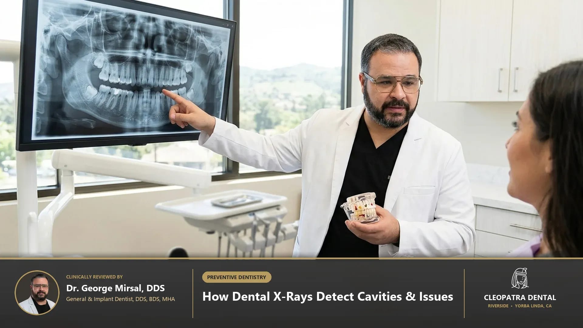
Patients researching how dental x-rays detect cavities and bone loss in Southern California deserve transparent, evidence-based guidance rooted in biological tooth preservation and long-term oral systemic health. At Cleopatra Dental, our general, restorative, and surgical clinicians evaluate every patient as a complete biomechanical system rather than treating isolated symptoms. In daily practice across Western Riverside County and North Orange County, we find that during a visual chairside exam, the contact points between adjacent back teeth, the roots embedded inside the jawbone, and the internal pulp chambers are completely hidden; digital X-ray photons pass differently through dense hydroxyapatite enamel (appearing bright white/radiopaque), softer dentin, and demineralized decay or bone infections (appearing dark/radiolucent)—exposing disease when only 30% mineral loss has occurred. This diagnostic precision prevents guesswork, minimizes chairside discomfort, and establishes predictable clinical outcomes for families and older adults alike. When digital radiographs reveal early horizontal or vertical bone loss, patients can schedule periodontal bone-level assessment and deep cleaning.
Modern dentistry has evolved rapidly away from aggressive mechanical preparation toward minimally invasive, biologically guided care. When evaluating types of dental x-rays bitewing periapical panoramic cbct, our diagnostic team examines how enamel thickness, underlying dentin tubules, periodontal ligament fibers, and occlusal biting forces interact over a 10- to 20-year horizon. By integrating digital optical scanning, low-dose imaging, and biocompatible materials coded under ADA CDT standards (D0274, D0220, D0330, D0367), we tailor each treatment plan to your specific anatomy, medical history, and comfort preferences. Our three-dimensional volumetric imaging also powers 3D CBCT-guided dental implant planning with sub-millimeter nerve safety.
Biological & Biomechanical Principles Behind types of dental x-rays bitewing periapical panoramic cbct
Every clinical decision in restorative and preventive dentistry depends on respecting microscopic tissue thresholds. Scientific studies confirm that staging interproximal caries radiographically (E1 outer half of enamel, E2 inner half of enamel, D1 outer third of dentin at the dentinoenamel junction [DEJ], D2 middle dentin, D3 deep pulp-approaching dentin) allows our clinicians to remineralize E1/E2 lesions non-invasively with 5% sodium fluoride varnish (D1206) while restoring D1/D2 lesions conservatively before root canal therapy is needed. When these quantitative parameters are carefully controlled at the chairside, natural dental tissues and prosthetic restorations withstand daily mastication forces—which routinely reach 70 to 150 pounds per square inch during normal chewing and exceed 250 pounds per square inch during nocturnal clenching—without premature wear, sensitivity, or structural fatigue. Curious about exact microsievert radiation numbers and pregnancy safety? Read our companion physics guide on the clinical truth about digital dental X-ray radiation doses and ALARA safety.
Advanced Biomaterials and Digital Diagnostic Standards
Material science plays an equally decisive role in clinical longevity and biocompatibility. In our clinical protocols, we utilize solid-state CMOS intraoral digital sensors (16-bit grayscale resolution with 20+ line pairs/mm), horizontal and vertical bitewing radiographs (CDT D0274), periapical radiographs (D0220 / D0230), full-mouth radiographic series (FMX, D0210), panoramic extraoral radiographs (D0330), and 0.1 mm voxel 3D Cone-Beam Computed Tomography (D0367). Unlike legacy dental materials that relied on mechanical wedging or high-expansion metals, contemporary ceramic, polymer, and biologic systems bond adhesively or integrate directly with host tissues. This seals out cariogenic oral bacteria, maintains neutral salivary pH balance, and delivers natural optical translucency that blends seamlessly with your surrounding smile. See how annual bitewings prevent emergency root canals in why 6-month dental checkups pair visual exams with preventive radiographs.
Side-by-Side Clinical Comparison & Candidacy Matrix
Choosing the right clinical pathway requires comparing treatment modalities across biological conservation, expected service life, patient comfort, and maintenance requirements. During your consultation, our doctors review high-resolution intraoral photographs with you on a chairside monitor so you can see exactly what we see. The clinical comparison matrix below summarizes how each diagnostic and therapeutic option performs in real-world practice. Discover how panoramic imaging uncovers asymptomatic jaw pathology in how dental X-rays and exams detect silent jaw cysts and systemic health markers.
| Digital Dental X-Ray Type (ADA CDT) | Anatomical Area Captured | Primary Oral Issues Diagnosed | Recommended Clinical Frequency (ADA Guidelines) |
|---|---|---|---|
| 4-Film Digital Bitewings (D0274) | Crowns of upper & lower back teeth + interdental alveolar bone crest | Interproximal "flossing-zone" cavities, overhanging fillings, subgingival tartar spurs, and early bone loss | Every 12 months (or 6–12 months for high cavity risk) |
| Periapical (PA) Radiograph (D0220 / D0230) | Entire single tooth from biting edge to root tip + surrounding jawbone | Root tip abscesses (periapical radiolucency), vertical root fractures, internal resorption, and root canal length | As needed for toothaches, crowns, or part of a 3–5 year Full-Mouth Series (D0210) |
| Panoramic Extraoral Radiograph (D0330) | Wide 2D sweep of entire upper/lower jaws, TMJ condyles, and maxillary sinuses | Impacted wisdom teeth, jaw cysts/tumors, supernumerary teeth in children, and carotid artery calcifications | Every 3 to 5 years or during wisdom tooth / orthodontic evaluation |
| 3D Cone-Beam CT Scan — CBCT (D0367) | 360-degree 3D volumetric reconstruction at 0.1 mm voxel accuracy | Exact 3D bone width for implants, hidden MB2 root canals, inferior alveolar nerve path, and sinus pathology | Targeted pre-surgical implant, complex root canal, or impacted extraction cases |
How We Customize Candidacy for Complex Medical Histories
No two mouths age or heal identically. Patients managing systemic conditions such as Type 2 diabetes, hypertension, autoimmune disorders, osteoporosis, or medication-induced dry mouth (xerostomia) require customized dental protocols. Before initiating treatment related to what do dental x-rays show, we review your current medications, allergies, and systemic health markers. Adjusting local anesthetic buffering, selecting hypoallergenic metal-free ceramics, and reinforcing salivary mineral buffering ensures safe, predictable healing for every age group. If you have existing restorations, learn how digital X-rays verify crown margins and detect recurrent decay.
Preventing Common Complications and Premature Failure
Even the finest dental restoration or preventive regimen requires protection against mechanical overload and bacterial biofilm accumulation. In clinical audits, the two leading causes of preventable dental breakdown are unaddressed nighttime teeth grinding (sleep bruxism) and interproximal plaque stagnation along the gumline. Addressing occlusal harmony early and establishing an easy-to-follow home care routine protects both your natural enamel and any restorative work for decades.
Long-Term Maintenance & Preventive Follow-Up in Southern California
Sustaining healthy clinical results over 10 to 20 years depends on partnering with a consistent dental team that monitors your gum attachment levels, bite balance, and restorative margins at every six-month checkup. At our Riverside and Yorba Linda locations, we combine ultrasonic hygiene care, digital bitewing monitoring, and occlusal nightguard checks to catch micro-changes long before they become costly emergencies.
2026 Cost Guide, ADA CDT Codes & PPO Insurance in Riverside and Yorba Linda
Financial clarity is a core pillar of patient trust at Cleopatra Dental. Treatment fees in Southern California vary according to diagnostic complexity, the number of teeth or quadrants involved, laboratory biomaterial selection, and your dental PPO insurance benefits. Before any procedure begins, our treatment coordinators verify your insurance eligibility and provide a clear, written breakdown organized by official American Dental Association Current Dental Terminology (CDT) codes.
Important Pricing Note: The prices shown in the table below are estimated ranges; to obtain accurate pricing tailored to your specific clinical needs and insurance coverage, please contact our Riverside or Yorba Linda office.
| Diagnostic Radiograph & ADA CDT Code | Estimated Cost & Price Range (2026) | Dental PPO Insurance Coverage & Diagnostic Value |
|---|---|---|
| Four Bitewing Radiographs — Digital CMOS (D0274) | $75 – $135 | Covered at 80% – 100% by PPO plans; catches cavities between teeth before they hurt |
| Intraoral Complete Series of Radiographs — FMX (D0210) | $145 – $245 | 18-to-20 digital views of every tooth and root; covered 80% – 100% every 3–5 years |
| Panoramic Radiographic Image (D0330) | $115 – $195 | Full jaw, wisdom teeth, and TMJ overview; covered 80% – 100% every 3–5 years |
| 3D Cone-Beam CT Capture & Interpretation (D0367) | $295 – $550 | Essential 3D roadmap for dental implants, sinus lifts, and hidden root canal anatomy |
Maximizing Dental PPO Insurance, HSA/FSA Funds & 0% Financing
Most PPO dental insurance plans categorize diagnostic exams, digital X-rays, and routine cleanings under preventive benefits covered at 80% to 100%, while basic and major restorative procedures receive 50% to 80% coverage up to your annual plan maximum (typically $1,500 to $2,500 per calendar year). For treatments that exceed annual insurance caps or involve elective cosmetic enhancements, patients can apply tax-free Health Savings Account (HSA) or Flexible Spending Account (FSA) dollars, or enroll in 0% APR promotional monthly financing through CareCredit, LendingClub, and Proceed Finance.
Step-by-Step Clinical Protocol & Long-Term Care Guidelines
Knowing exactly what happens before, during, and after your visit eliminates dental anxiety and empowers you to participate actively in your oral health. Our clinical workflow enforces co-reviewed by supervising dentists Dr. George Mirsal, DDS and Dr. Engy Halim, DDS alongside Registered Dental Hygienist JoAnne Schram, RDH per California B&P Code § 1907–1926: reviewing your enlarged digital radiographs together on a 4K chairside monitor so you see every interproximal contact, bone crest level, and root tip with complete transparency. By breaking complex care into calm, well-paced stages, we ensure your comfort from initial check-in through long-term maintenance. Instant chairside CMOS digital radiography, iTero NIRI caries scanning, and 3D CBCT imaging are available at Cleopatra Dental in Yorba Linda, CA.
Step 1: Comprehensive Digital Assessment and Shade/Bite Mapping
Your appointment begins with a conversation about your goals, symptoms, and past dental experiences, followed by gentle digital diagnostics. Using radiation-free iTero 3D optical scanning, intraoral high-definition cameras, and ultra-low-dose digital radiographs when indicated, we map your teeth, gums, and bite registration within minutes—eliminating messy impression trays and providing an immediate 3D visual roadmap of your mouth. Inland Empire patients receive identical high-resolution digital diagnostic imaging at our Riverside diagnostic and family dental center.
Step 2: Precision Chairside Execution and Post-Care Verification
During active care, we prioritize profound comfort using topical numbing gels, pH-buffered local anesthetics, and optional nitrous oxide or oral conscious sedation. Every margin, contact point, and biting surface is verified under high magnification before you leave the chair. You also receive written post-visit care instructions and a direct follow-up call from our clinical team to confirm your recovery is progressing smoothly.
Frequently Asked Questions About How Digital Dental X-Rays Diagnose Hidden Cavities & Bone Loss
How does a cavity show up on a digital dental X-ray?
Healthy tooth enamel is the hardest, most mineral-dense substance in the human body, so it blocks X-ray photons and appears bright white on the screen. When cavity-causing bacteria produce acid that dissolves mineral out of the enamel between two teeth, that softer area lets more photons pass through—showing up as a distinct dark V-shaped shadow right at the contact point.
Can dental X-rays detect gum disease and bone loss?
Yes! While a periodontal probe measures gum pocket depth, digital bitewing and periapical X-rays show the exact height and density of the interdental jawbone supporting your teeth. Normally, the bone crest sits 1.0 to 2.0 millimeters below the enamel edge; horizontal or vertical dips in that bone line reveal active or past periodontal disease.
Why can’t a panoramic X-ray replace annual bitewing X-rays for finding cavities?
A panoramic X-ray (CDT D0330) rotates outside your head to capture a wide view of your wisdom teeth, jaw joints, and sinuses, which causes slight overlapping of the premolars and molars. Intraoral digital bitewings (D0274) sit directly behind your teeth at a perpendicular angle, providing 5x sharper micro-resolution to spot tiny early cavities between teeth.
What can a 3D CBCT scan find that a regular 2D dental X-ray misses?
A regular 2D X-ray compresses a three-dimensional tooth into a flat shadow, which can hide an infection sitting directly behind a dense root or conceal a thin buccal bone wall. A 3D CBCT scan (CDT D0367) lets our doctors view cross-sectional slices from every angle—revealing hidden fourth root canals, hairline root fractures, sinus infections of dental origin, and exact millimeter bone thickness for implants.
Verified Branch & Location: Visit Cleopatra Dental in Yorba Linda at 18651 Yorba Linda Blvd, Yorba Linda, CA 92886 (serving East Lake Village, Travis Ranch, Bryant Ranch, Kerrigan Ranch, Anaheim Hills, and Placentia near Yorba Linda Blvd and Imperial Hwy), or view directions and verified patient reviews on Cleopatra Dental Yorba Linda digital dental X-ray and 3D CBCT diagnostic center on Google Maps.
Clinically & Technically Reviewed By:
Dr. George Mirsal, DDS, BDS, MHA — General & Implant Dentist
Dr. Engy Halim, DDS, BDS — General & Cosmetic Dentist
JoAnne Schram, RDH, BVoc, MEA — Registered Dental Hygienist (Supervised by DDS per CA B&P § 1907–1926)
Medical Disclaimer: This content is clinically reviewed for educational purposes in accordance with American Dental Association (ADA) and Dental Board of California guidelines and does not replace a direct in-person dental examination.