PRF Membrane Graft Step-by-Step: Accelerated Dental Implant Healing
October 26, 2026
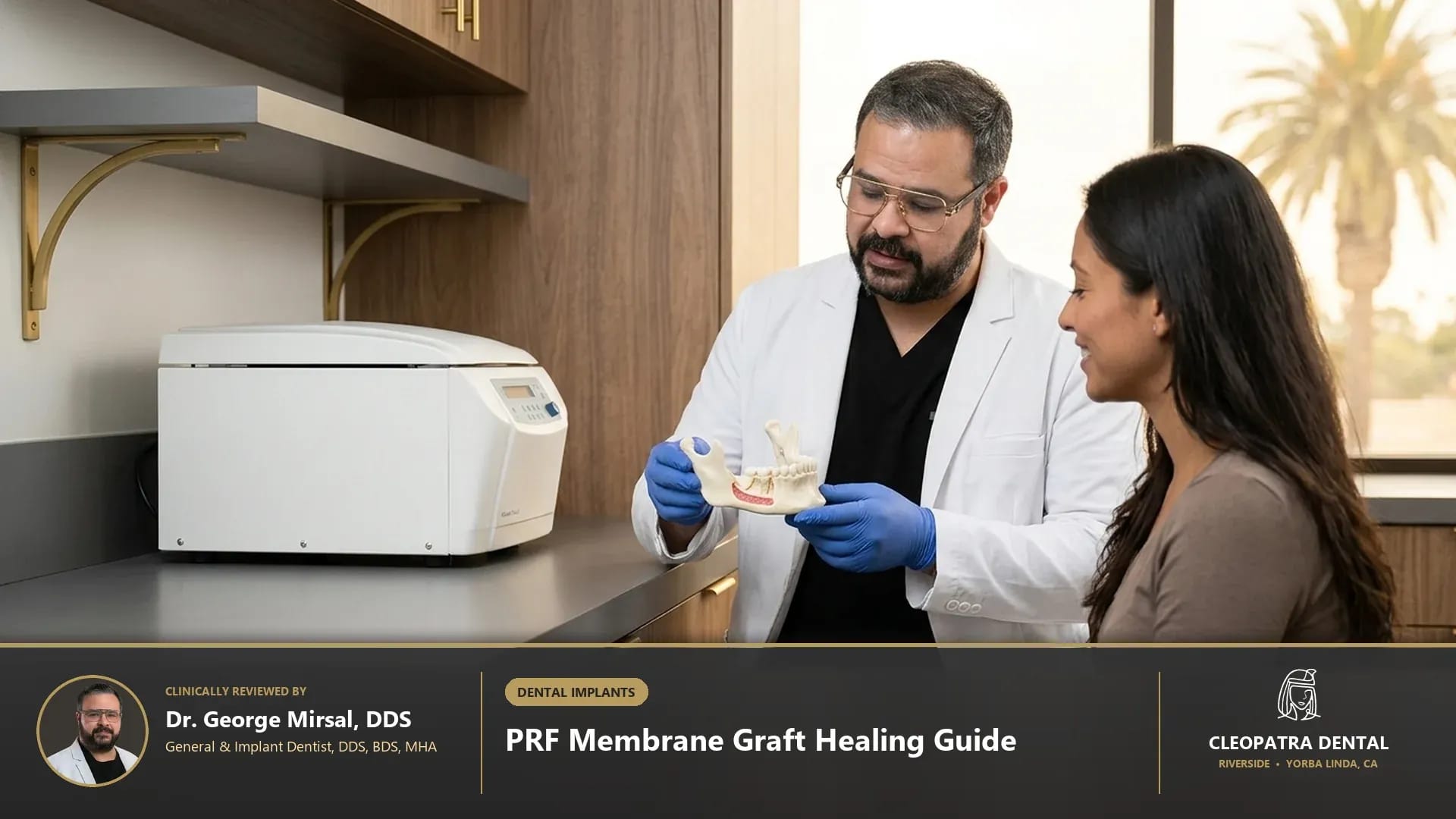
Quick Verified Summary: Autologous Leukocyte- and Platelet-Rich Fibrin (L-PRF) membrane grafting in Riverside and Yorba Linda concentrates your own blood platelets, leukocytes, and fibrin matrix via anticoagulant-free centrifugation at 2,700 RPM for 12 minutes. Placed into extraction sockets or bone grafts (CDT D7953), PRF releases growth factors for 7 to 14 days, cutting soft-tissue healing time in half.
Clinical Foundations: Understanding PRF Membrane Graft Step-by-Step: Accelerated Dental Implant Healing
Patients researching prf membrane graft step by step in Southern California deserve transparent, evidence-based guidance rooted in biological tooth preservation and long-term oral systemic health. At Cleopatra Dental, our general, restorative, and surgical clinicians evaluate every patient as a complete biomechanical system rather than treating isolated symptoms. In daily practice across Western Riverside County and North Orange County, we find that pre-operative 3D CBCT bone density evaluation (CDT D0367) identifies buccal plate dehiscences, fenestrations, and maxillary sinus floor proximity requiring guided bone regeneration alongside autologous fibrin biologics. This diagnostic precision prevents guesswork, minimizes chairside discomfort, and establishes predictable clinical outcomes for families and older adults alike. Combining autologous fibrin matrices with biologically enhanced dental implant surgery maximizes bone-to-implant contact in compromised ridges.
Modern dentistry has evolved rapidly away from aggressive mechanical preparation toward minimally invasive, biologically guided care. When evaluating prf membrane graft procedure steps, our diagnostic team examines how enamel thickness, underlying dentin tubules, periodontal ligament fibers, and occlusal biting forces interact over a 10- to 20-year horizon. By integrating digital optical scanning, low-dose imaging, and biocompatible materials coded under ADA CDT standards (D7953, D7950, D6010, D7210), we tailor each treatment plan to your specific anatomy, medical history, and comfort preferences. We routinely place L-PRF membranes immediately following atraumatic tooth extraction and ridge preservation to prevent post-extraction alveolar collapse.
Biological & Biomechanical Principles Behind prf membrane graft procedure steps
Every clinical decision in restorative and preventive dentistry depends on respecting microscopic tissue thresholds. Scientific studies confirm that drawing 20 to 40 mL of peripheral venous blood into additive-free glass-coated tubes and spinning immediately at 2,700 RPM (approximately 400 to 700 RCF) for 12 minutes polymerizes a three-dimensional trimolecular fibrin scaffold trapping 95% of platelets and 70% of leukocytes. When these quantitative parameters are carefully controlled at the chairside, natural dental tissues and prosthetic restorations withstand daily mastication forces—which routinely reach 70 to 150 pounds per square inch during normal chewing and exceed 250 pounds per square inch during nocturnal clenching—without premature wear, sensitivity, or structural fatigue. See how biological membranes integrate into our week-by-week dental implant surgery preparation and recovery timeline.
Advanced Biomaterials and Digital Diagnostic Standards
Material science plays an equally decisive role in clinical longevity and biocompatibility. In our clinical protocols, we utilize 100% autologous Leukocyte-Platelet-Rich Fibrin (L-PRF) membranes and liquid injectable i-PRF combined with mineralized freeze-dried bone allograft (FDBA) or deproteinized bovine bone mineral (CDT D7953) to form cohesive sticky bone that supports 35 to 45 Ncm implant insertion torque, an Implant Stability Quotient (ISQ) above 70, and definitive monolithic zirconia crowns. Unlike legacy dental materials that relied on mechanical wedging or high-expansion metals, contemporary ceramic, polymer, and biologic systems bond adhesively or integrate directly with host tissues. This seals out cariogenic oral bacteria, maintains neutral salivary pH balance, and delivers natural optical translucency that blends seamlessly with your surrounding smile. Explore how regenerated alveolar bone supports different fixtures in our guide to the main types of titanium and zirconia dental implants.
Side-by-Side Clinical Comparison & Candidacy Matrix
Choosing the right clinical pathway requires comparing treatment modalities across biological conservation, expected service life, patient comfort, and maintenance requirements. During your consultation, our doctors review high-resolution intraoral photographs with you on a chairside monitor so you can see exactly what we see. The clinical comparison matrix below summarizes how each diagnostic and therapeutic option performs in real-world practice. If you are weighing endodontic salvage against removal and socket grafting, read when surgical tooth extraction is safer than saving a damaged tooth.
| Biologic Parameter | Second-Generation L-PRF Membrane | First-Generation PRP (Platelet-Rich Plasma) | Synthetic Collagen Resorbable Membrane |
|---|---|---|---|
| Biochemical Composition | 100% autologous blood; zero bovine thrombin or anticoagulants | Requires sodium citrate anticoagulant and bovine thrombin/calcium chloride | Cross-linked porcine or bovine Type I/III pericardium collagen |
| Growth Factor Release Kinetics | Slow, sustained cytokine release over 7 to 14 days during fibrin remodeling | Rapid burst release of 80%+ growth factors within first 12 to 24 hours | Zero intrinsic growth factors; acts strictly as a passive barrier |
| Immunological & Leukocyte Activity | Enriched with host leukocytes and monocytes that resist oral bacteria | Low or variable leukocyte concentration depending on double-spin protocol | Foreign-body enzymatic resorption over 8 to 24 weeks |
| Open-Wound Exposure Tolerance | Epithelializes rapidly within 5–7 days even when left partially exposed | Liquid/gel consistency cannot serve as a suturable barrier membrane | Premature exposure often leads to bacterial contamination and graft loss |
| Chairside Preparation Time | Single 12-minute spin (2,700 RPM) during local anesthesia onset | Complex 30-minute double-centrifugation protocol with chemical additives | Ready out of sterile package but adds significant material cost ($300–$600) |
How We Customize Candidacy for Complex Medical Histories
No two mouths age or heal identically. Patients managing systemic conditions such as Type 2 diabetes, hypertension, autoimmune disorders, osteoporosis, or medication-induced dry mouth (xerostomia) require customized dental protocols. Before initiating treatment related to prf membrane graft how to prepare, we review your current medications, allergies, and systemic health markers. Adjusting local anesthetic buffering, selecting hypoallergenic metal-free ceramics, and reinforcing salivary mineral buffering ensures safe, predictable healing for every age group.
Preventing Common Complications and Premature Failure
Even the finest dental restoration or preventive regimen requires protection against mechanical overload and bacterial biofilm accumulation. In clinical audits, the two leading causes of preventable dental breakdown are unaddressed nighttime teeth grinding (sleep bruxism) and interproximal plaque stagnation along the gumline. Addressing occlusal harmony early and establishing an easy-to-follow home care routine protects both your natural enamel and any restorative work for decades.
Long-Term Maintenance & Preventive Follow-Up in Southern California
Sustaining healthy clinical results over 10 to 20 years depends on partnering with a consistent dental team that monitors your gum attachment levels, bite balance, and restorative margins at every six-month checkup. At our Riverside and Yorba Linda locations, we combine ultrasonic hygiene care, digital bitewing monitoring, and occlusal nightguard checks to catch micro-changes long before they become costly emergencies.
2026 Cost Guide, ADA CDT Codes & PPO Insurance in Riverside and Yorba Linda
Financial clarity is a core pillar of patient trust at Cleopatra Dental. Treatment fees in Southern California vary according to diagnostic complexity, the number of teeth or quadrants involved, laboratory biomaterial selection, and your dental PPO insurance benefits. Before any procedure begins, our treatment coordinators verify your insurance eligibility and provide a clear, written breakdown organized by official American Dental Association Current Dental Terminology (CDT) codes.
Important Pricing Note: The prices shown in the table below are estimated ranges; to obtain accurate pricing tailored to your specific clinical needs and insurance coverage, please contact our Riverside or Yorba Linda office.
| Regenerative Procedure & ADA CDT Code | Estimated Cost & Price Range (2026) | Clinical Application & Insurance Coverage Notes |
|---|---|---|
| Autologous PRF Collection & Membrane Fabrication (D7921) | $250 – $550 | Includes chairside phlebotomy and centrifugation of 2 to 4 L-PRF clots/membranes |
| Bone Replacement Graft for Ridge Preservation (D7953) | $450 – $950 | Particulate mineralized bone graft hydrated with liquid PRF inside fresh extraction socket |
| Guided Tissue Regeneration Barrier Membrane (D4266 / D7956) | $350 – $750 | Layered L-PRF membrane closure over particulate graft; often bundled in implant packages |
| Sinus Augmentation with Lateral or Crestal PRF Approach (D7951 / D7952) | $1,200 – $2,600 | Elevating Schneiderian membrane cushioned with L-PRF matrices prior to posterior maxillary implants |
| Surgical Extraction + Socket Graft + PRF Turnkey Bundle | $950 – $1,650 | Comprehensive pre-implant site preservation eligible for PPO oral surgery benefits and CareCredit |
Maximizing Dental PPO Insurance, HSA/FSA Funds & 0% Financing
Most PPO dental insurance plans categorize diagnostic exams, digital X-rays, and routine cleanings under preventive benefits covered at 80% to 100%, while basic and major restorative procedures receive 50% to 80% coverage up to your annual plan maximum (typically $1,500 to $2,500 per calendar year). For treatments that exceed annual insurance caps or involve elective cosmetic enhancements, patients can apply tax-free Health Savings Account (HSA) or Flexible Spending Account (FSA) dollars, or enroll in 0% APR promotional monthly financing through CareCredit, LendingClub, and Proceed Finance.
Step-by-Step Clinical Protocol & Long-Term Care Guidelines
Knowing exactly what happens before, during, and after your visit eliminates dental anxiety and empowers you to participate actively in your oral health. Our clinical workflow enforces sustained 7-to-14-day release of Platelet-Derived Growth Factor (PDGF-AB), Transforming Growth Factor Beta-1 (TGF-B1), and Vascular Endothelial Growth Factor (VEGF) stimulates angiogenesis, osteoblast chemotaxis, and reduces alveolar osteitis (dry socket) incidence by over 80%. By breaking complex care into calm, well-paced stages, we ensure your comfort from initial check-in through long-term maintenance. Chairside venipuncture and FDA-cleared horizontal centrifugation are performed in-house at our Riverside oral surgery and implant center.
Step 1: Comprehensive Digital Assessment and Shade/Bite Mapping
Your appointment begins with a conversation about your goals, symptoms, and past dental experiences, followed by gentle digital diagnostics. Using radiation-free iTero 3D optical scanning, intraoral high-definition cameras, and ultra-low-dose digital radiographs when indicated, we map your teeth, gums, and bite registration within minutes—eliminating messy impression trays and providing an immediate 3D visual roadmap of your mouth. Patients in Orange County can also benefit from autologous L-PRF bone and soft-tissue grafting at Cleopatra Dental in Yorba Linda, CA.
Step 2: Precision Chairside Execution and Post-Care Verification
During active care, we prioritize profound comfort using topical numbing gels, pH-buffered local anesthetics, and optional nitrous oxide or oral conscious sedation. Every margin, contact point, and biting surface is verified under high magnification before you leave the chair. You also receive written post-visit care instructions and a direct follow-up call from our clinical team to confirm your recovery is progressing smoothly.
Frequently Asked Questions About PRF Membrane Graft Step-by-Step: Accelerated Dental Implant Healing
Is the PRF blood draw painful or risky for dental patients?
No. Obtaining blood for Platelet-Rich Fibrin is identical to a routine lab blood test, requiring only 2 to 4 small vials drawn from an arm vein while your local dental anesthesia takes effect. Because L-PRF is 100% derived from your own blood without chemical additives or animal enzymes, there is zero risk of allergy, disease transmission, or immune rejection.
How do I prepare my body in the 24 hours before a PRF dental procedure?
Hydration is the single most important preparation step: drink 64 to 80 ounces of water the day before and the morning of your appointment so venous access is effortless and your plasma volume produces thick, nutrient-dense fibrin membranes. Avoid alcohol and high-fat meals for 24 hours prior to surgery.
Does PRF really prevent dry socket after wisdom tooth or molar extractions?
Yes. Clinical trials published in oral and maxillofacial surgery journals demonstrate that sealing extraction sockets with autologous L-PRF clots reduces dry socket (alveolar osteitis) rates by more than 80% while significantly lowering post-operative pain scores on days 1 through 5.
Can PRF shorten the waiting time between tooth extraction and dental implant placement?
By accelerating angiogenesis and woven bone trabecular density through sustained 14-day release of PDGF and VEGF growth factors, sticky bone grafts enriched with L-PRF typically reach implant-ready radiographic density in 10 to 12 weeks instead of the conventional 16 to 24 weeks.
Verified Branch & Location: Visit Cleopatra Dental in Riverside at 2955 Van Buren Boulevard, Ste H4, Riverside, CA 92503 (serving Arlington, La Sierra, Canyon Crest, Wood Streets, Orangecrest, and Corona near Van Buren Blvd and CA-91), or view directions and verified patient reviews on Cleopatra Dental Riverside PRF & bone grafting clinic on Google Maps.
Clinically & Technically Reviewed By:
Dr. George Mirsal, DDS, BDS, MHA — General & Implant Dentist
Medical Disclaimer: This content is clinically reviewed for educational purposes in accordance with American Dental Association (ADA) and Dental Board of California guidelines and does not replace a direct in-person dental examination.